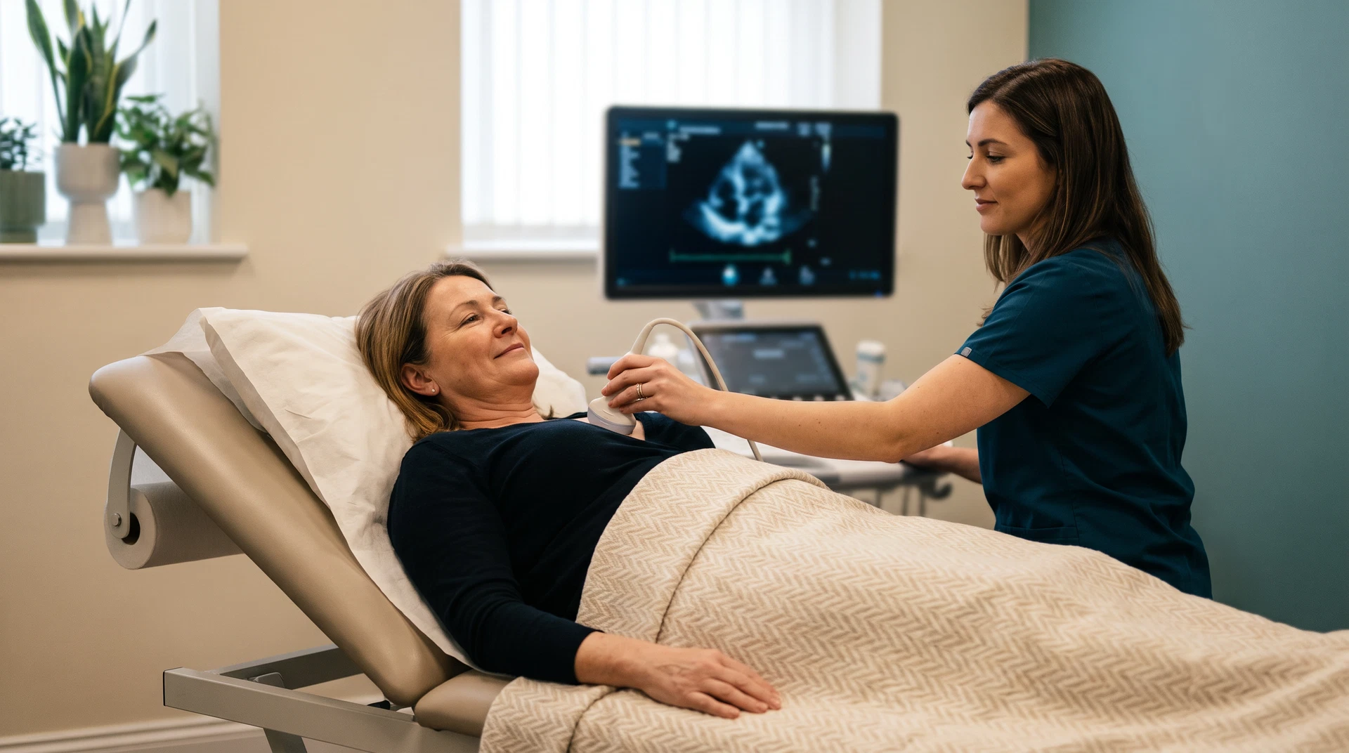
An echocardiogram (echo) uses ultrasound waves to create images of the heart. It can assess cardiac structure, pumping function, valves and blood flow, but the meaning of any finding depends on your symptoms, examination and wider clinical history.[1]
Before Your Appointment
- Wear clothing that is easy to remove from the upper body; a gown may be provided
- For many standard transthoracic echocardiograms, normal eating and drinking may be possible; other types of echo can have different instructions
- Continue prescribed medicines unless the clinic or referring clinician gives different advice
- Bring a list of current medicines and relevant previous cardiac test information if requested
- Tell the clinic about access, privacy or chaperone needs before the appointment
- The clinic confirms preparation, expected appointment length, aftercare and any activity advice for the selected pathway at booking
During the Scan
You will lie on your left side on an examination couch. A clinician applies gel to your chest and moves a probe across different positions to obtain images of your heart. You may hear a "whooshing" Doppler signal. Ultrasound is generally well tolerated. Some examinations can cause temporary pressure or discomfort depending on the area examined. It does not use ionising radiation.[3]
What the Scan Assesses
| Structure | What We Measure | Clinical Significance |
|---|---|---|
| Left ventricle | Size, wall thickness, ejection fraction (pumping function) | Heart failure, cardiomyopathy, hypertensive heart disease |
| Heart valves | Opening, closing, regurgitation (leaking), stenosis (narrowing) | Valvular heart disease — aortic stenosis, mitral regurgitation |
| Right heart | Size, pressure estimates, tricuspid valve function | Pulmonary hypertension, right heart failure |
| Pericardium | Fluid around the heart (effusion) | Pericarditis, post-surgical changes |
| Aorta | Root diameter, ascending aorta size | Aortic aneurysm, Marfan syndrome |
| Blood flow | Doppler velocities across all valves | Severity grading of valve disease, diastolic function |
After the Scan
The clinic explains the reporting process and appropriate follow-up route for your examination at booking. If a finding needs further assessment, the next step depends on the clinical context and the relevant specialist or urgent-care pathway.
What to expect
Tell the clinician if you are uncomfortable or need to pause. The team can explain each step and discuss questions about the scan.
Frequently Asked Questions
Is an echocardiogram uncomfortable?
Ultrasound is generally well tolerated. Some examinations can cause temporary pressure or discomfort depending on the area examined.
How long does it take?
The clinic confirms the expected appointment length for your pathway at booking.
Do I need a referral?
The clinic confirms the appropriate appointment route and any referral requirements for your echocardiogram pathway at booking.
Is there any radiation?
No. Echocardiography uses ultrasound waves rather than ionising radiation.
When will I get results?
The clinic explains the reporting process and appropriate follow-up route for your examination at booking.
Can I eat and drink before the scan?
The clinic will confirm any preparation instructions for your examination at booking.
Related Guides
ECG vs Echocardiogram
What's the difference?
When to See a Cardiologist
Warning signs explained
Heart Palpitations
Causes and tests
Echocardiogram Service
Book your heart scan
References
- [1] British Society of Echocardiography. A minimum dataset for a standard adult transthoracic echocardiogram. 2020. View source
- [2] Marwick TH. The role of echocardiography in heart failure. J Nucl Med. 2015;56(Suppl 4):31S–38S. View source
- [3] Cheitlin MD, et al. ACC/AHA guidelines for the clinical application of echocardiography. Circulation. 1997;95(6):1686–1744. View source
- [4] NICE CG187. Acute heart failure: diagnosis and management. 2014. View source
- [5] Lancellotti P, et al. European Association of Echocardiography recommendations for the assessment of valvular regurgitation. Eur J Echocardiogr. 2010;11(4):307–332. View source
- [6] Nagueh SF, et al. Recommendations for the evaluation of left ventricular diastolic function by echocardiography. J Am Soc Echocardiogr. 2016;29(4):277–314. View source